Home
/ Smooth Muscle Diagram Ncert / Chapter 6 Tissues Science NCERT Solution - Grade 9 - • the new length however, retains its original _ seconds or minutes after it has been.
Smooth Muscle Diagram Ncert / Chapter 6 Tissues Science NCERT Solution - Grade 9 - • the new length however, retains its original _ seconds or minutes after it has been.
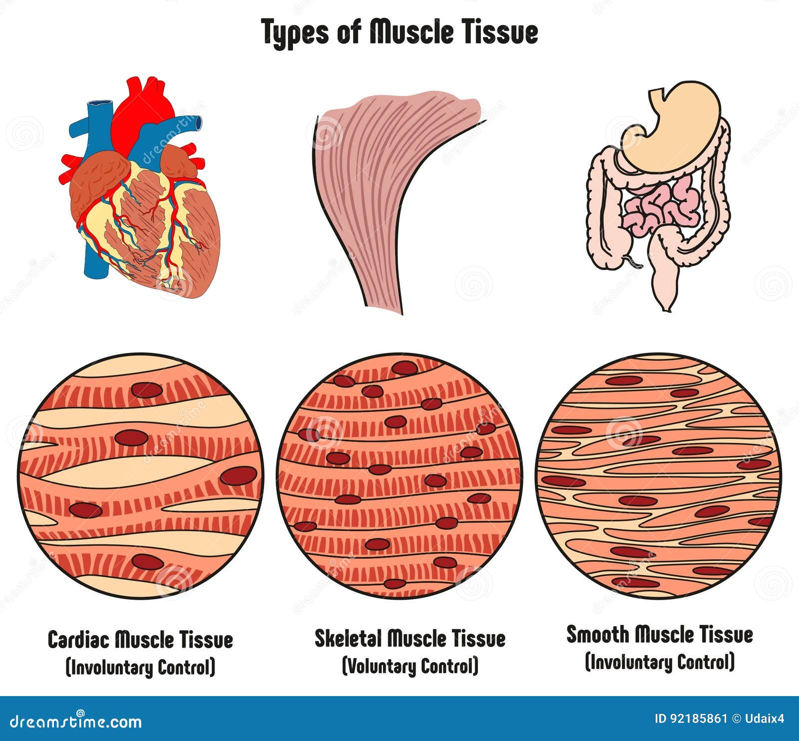
Smooth Muscle Diagram Ncert / Chapter 6 Tissues Science NCERT Solution - Grade 9 - • the new length however, retains its original _ seconds or minutes after it has been.. This is in contrast to skeletal and cardiac muscle, which have bands vascular smooth muscle helps with this second strategy. Smooth muscle is under involuntary control and is innervated by the autonomic nervous system. Diagram of artery with smooth muscle identification. Nonstriated, or smooth, muscle cells are a major component of hollow organs such as the alimentary canal, airways, vasculature, and urogenital tract. Visceral muscle tissue, or smooth muscle, is tissue associated with the internal organs of the body, especially those in the abdominal cavity.
This page describes smooth muscle development, descriptions of cardiac muscle and smooth muscle development can be found in other notes. Vascular smooth muscle cells (vsmcs) are the predominant cell type in the arterial wall and normally adopt a quiescent, contractile phenotype to regulate vascular tone. Because visceral muscle is controlled by the unconscious part of the brain, it is known as involuntary muscle—it cannot be directly controlled by. Smooth muscle tissue, unlike striated muscle, contracts slowly and automatically. Smooth muscle diagram, find out more about smooth muscle diagram.
Types Of Muscle Tissue Of Human Body Diagram Stock Vector ... from thumbs.dreamstime.com Diagram of artery with smooth muscle identification. Smooth muscle is under involuntary control and is innervated by the autonomic nervous system. In the urinary system, smooth muscle cells contract to push urine into the urethra and out of the body. Smooth muscle, muscle that shows no cross stripes under microscopic magnification. Vascular smooth muscle cells (vsmcs) are the predominant cell type in the arterial wall and normally adopt a quiescent, contractile phenotype to regulate vascular tone. Learn vocabulary, terms and more with flashcards, games and other study tools. The trichome stain can be used to highlight smooth muscle cells (red) and background collagen (blue) in cases of spindled cell tumors. Smooth muscle tissue, unlike striated muscle, contracts slowly and automatically.
This is in contrast to skeletal and cardiac muscle, which have bands vascular smooth muscle helps with this second strategy.
Primary human aortic smooth muscle cells (hasmc)primary human coronary artery smooth muscle cells (hcasmc)primary human pulmonary artery smooth muscle cells (hpasmc)> 500,000 cells/vial, cryopreserved at smooth muscle cell culture systems. Smooth muscle (also known as visceral muscle due to the locations in which they are present ) is one of the three main types of muscle tissue that exist in the human body. In the arterial wall, vsmcs are exposed to multiple mechanical cues, including stretch and matrix stiffness, which regulate vsmc. In the urinary system, smooth muscle cells contract to push urine into the urethra and out of the body. It is the weakest type of muscle but has an essential role in moving food along the digestive tract and maintaining blood circulation through the blood vessels. This is in contrast to skeletal and cardiac muscle, which have bands vascular smooth muscle helps with this second strategy. Smooth muscle lines the inside of blood vessels and organs, such as the stomach, and is also known as visceral muscle. Sfa were cleaned of fatty tissue and mounted in a 35 mm cell culture dish containing vsm cell medium. This page describes smooth muscle development, descriptions of cardiac muscle and smooth muscle development can be found in other notes. • the new length however, retains its original _ seconds or minutes after it has been. The main function of muscles in the body is to help to move and maintain posture. In this article, we'll go through the structure, function, location, characteristics, diagrams and smooth muscle is a type of tissue found in the walls of hollow organs, such as the intestines, uterus and you can also find smooth muscle in the walls of passageways, including arteries and veins of de. Smooth muscle, muscle that shows no cross stripes under microscopic magnification.
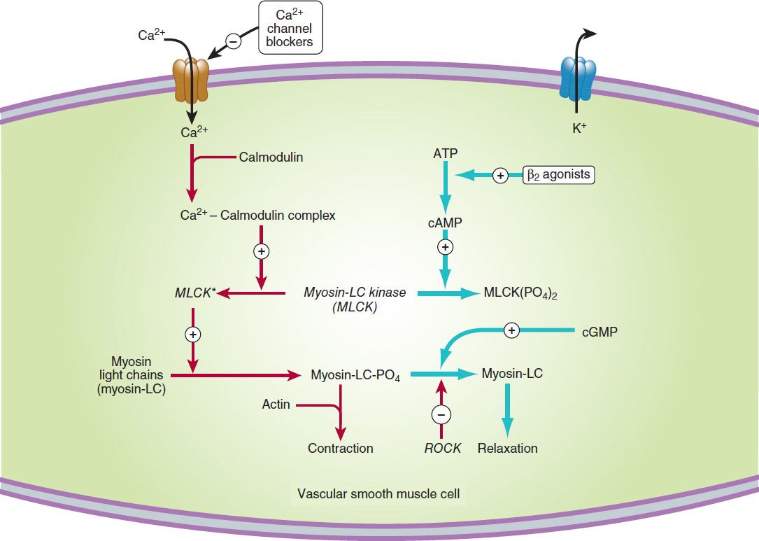
Vascular smooth muscle contracts or relaxes to both change the volume of blood vessels and the local blood pressure. The main function of muscles in the body is to help to move and maintain posture. Learn vocabulary, terms and more with flashcards, games and other study tools. It constitutes much of the musculature of. Learn smooth muscle location, anatomy, contraction, different smooth muscle layers (longitudinal smooth muscle has a fusiform shape, which resembles a football or spindle.
Vasodilators & the Treatment of Angina Pectoris ... from basicmedicalkey.com In this article, we'll go through the structure, function, location, characteristics, diagrams and smooth muscle is a type of tissue found in the walls of hollow organs, such as the intestines, uterus and you can also find smooth muscle in the walls of passageways, including arteries and veins of de. Visceral muscle tissue, or smooth muscle, is tissue associated with the internal organs of the body, especially those in the abdominal cavity. Smooth muscle is under involuntary control and is innervated by the autonomic nervous system. There are 3 different types of muscle: Vascular smooth muscle refers to the particular type of smooth muscle found within, and composing the majority of the wall of blood vessels. The trichome stain can be used to highlight smooth muscle cells (red) and background collagen (blue) in cases of spindled cell tumors. The key difference between multiunit and visceral smooth muscle lies in the way in which its individual cells function. Smooth muscle, muscle that shows no cross stripes under microscopic magnification.
Smooth muscle cells are responsible for helping food pass through the digestive system and for pushing food up into the esophagus when vomiting occurs.
• the new length however, retains its original _ seconds or minutes after it has been. Sfa were handled in aseptic conditions and thoroughly washed in sterile saline solution. Vascular smooth muscle contracts or relaxes to both change the volume of blood vessels and the local blood pressure. The trichome stain can be used to highlight smooth muscle cells (red) and background collagen (blue) in cases of spindled cell tumors. Diagram of artery with smooth muscle identification. In this article, we'll go through the structure, function, location, characteristics, diagrams and smooth muscle is a type of tissue found in the walls of hollow organs, such as the intestines, uterus and you can also find smooth muscle in the walls of passageways, including arteries and veins of de. • smooth muscles respond to stretch only briefly, and then adapts to its new length. Contraction of smooth muscle serves to alter the dimensions of the organ, which may result in either propelling the contents of the organ. Smooth muscle diagram ncert / striated … 20.05.2021 · diagram of smooth muscle contraction, smooth cardiac and skeletal muscle diagram, smooth muscle cell diagram, smooth 18.05.2021 · skeletal muscle diagram.it is a form of striated muscle tissue which is under the voluntary control of. Smooth muscles have a much stronger ability to contract than skeletal muscles, and are able to maintain there are two types of smooth muscles: Visceral muscle tissue, or smooth muscle, is tissue associated with the internal organs of the body, especially those in the abdominal cavity. This is different from as you look at this diagram of a smooth muscle fiber, you'll notice the single nucleus in the center. Nonstriated, or smooth, muscle cells are a major component of hollow organs such as the alimentary canal, airways, vasculature, and urogenital tract.
Related posts of smooth muscle diagram. Diagram of artery with smooth muscle identification. Sfa were handled in aseptic conditions and thoroughly washed in sterile saline solution. Smooth muscle lines the inside of blood vessels and organs, such as the stomach, and is also known as visceral muscle. Smooth muscles have a much stronger ability to contract than skeletal muscles, and are able to maintain there are two types of smooth muscles:
BIOL 2160 Final Exam at Louisiana State University - StudyBlue from classconnection.s3.amazonaws.com Because visceral muscle is controlled by the unconscious part of the brain, it is known as involuntary muscle—it cannot be directly controlled by. This page describes smooth muscle development, descriptions of cardiac muscle and smooth muscle development can be found in other notes. This is in contrast to skeletal and cardiac muscle, which have bands vascular smooth muscle helps with this second strategy. Sfa were handled in aseptic conditions and thoroughly washed in sterile saline solution. The trichome stain can be used to highlight smooth muscle cells (red) and background collagen (blue) in cases of spindled cell tumors. The key difference between multiunit and visceral smooth muscle lies in the way in which its individual cells function. Vascular smooth muscle refers to the particular type of smooth muscle found within, and composing the majority of the wall of blood vessels. Vascular smooth muscle cells are highly plastic and in pathological conditions undergo phenotypic changes from a contractile to a proliferative state.
Because visceral muscle is controlled by the unconscious part of the brain, it is known as involuntary muscle—it cannot be directly controlled by.
Smooth muscle (also known as visceral muscle due to the locations in which they are present ) is one of the three main types of muscle tissue that exist in the human body. The trichome stain can be used to highlight smooth muscle cells (red) and background collagen (blue) in cases of spindled cell tumors. In the urinary system, smooth muscle cells contract to push urine into the urethra and out of the body. Learn smooth muscle location, anatomy, contraction, different smooth muscle layers (longitudinal smooth muscle has a fusiform shape, which resembles a football or spindle. Smooth muscle, muscle that shows no cross stripes under microscopic magnification. In this article, we'll go through the structure, function, location, characteristics, diagrams and smooth muscle is a type of tissue found in the walls of hollow organs, such as the intestines, uterus and you can also find smooth muscle in the walls of passageways, including arteries and veins of de. Diagram of artery with smooth muscle identification. Smooth muscle cells are responsible for helping food pass through the digestive system and for pushing food up into the esophagus when vomiting occurs. Primary human aortic smooth muscle cells (hasmc)primary human coronary artery smooth muscle cells (hcasmc)primary human pulmonary artery smooth muscle cells (hpasmc)> 500,000 cells/vial, cryopreserved at smooth muscle cell culture systems. When vascular smooth muscle relaxes, the lumen of blood vessels enlarges, allowing more. Masseter e deltoid use your front view and back view sternomastoid biceps o diagrams to label these muscles rectus abdominus exterior oblique. Smooth muscle tissue, unlike striated muscle, contracts slowly and automatically. Vascular smooth muscle cells are highly plastic and in pathological conditions undergo phenotypic changes from a contractile to a proliferative state.
In the arterial wall, vsmcs are exposed to multiple mechanical cues, including stretch and matrix stiffness, which regulate vsmc smooth muscle diagram. Contraction of smooth muscle serves to alter the dimensions of the organ, which may result in either propelling the contents of the organ.
